Person undergoing a CAT scan in hospital. PET scan equipment. Medical CT scan of patient.
Browse 4,800+ pet scan stock photos and images available, or search for pet scan prostate or pet scan alzheimer to find more great stock photos and pictures.
Person undergoing a CAT scan in hospital. PET scan equipment. Medical CT scan of patient.
Pet tracking line icons. Vector illustration include icon - trace, collar, microchip, magnifier, battery, dog, cat tracking outline pictogram for animal search. 64x64 Pixel Perfect, Editable Stroke.

Doctor getting a patient ready for a medical scan at the hospital - healthcare and medicine concepts
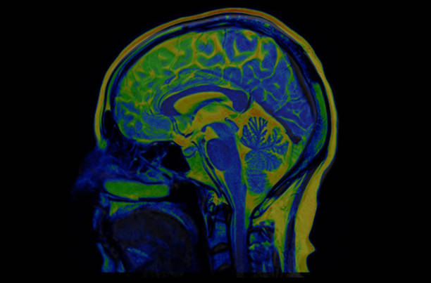
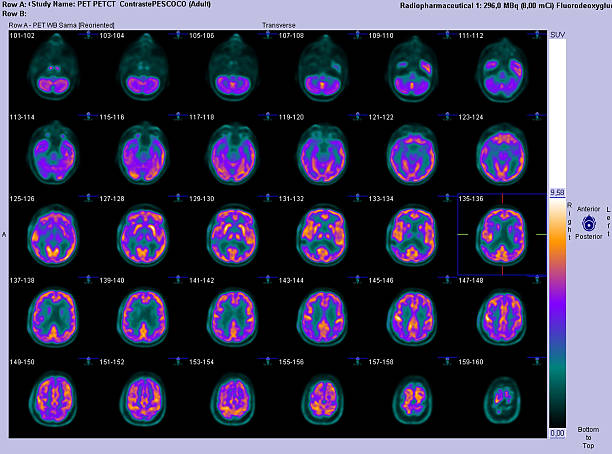
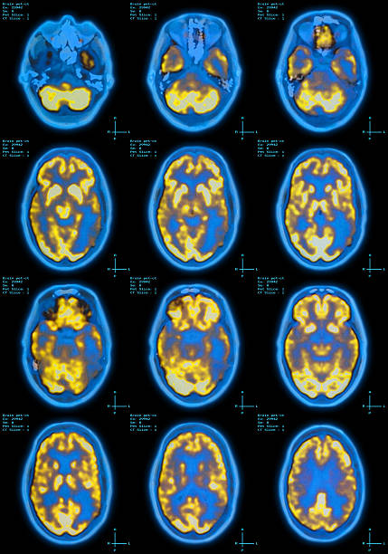
These are computer generated images called PET (positron emission tomography) of the brain. It consists of injecting a radioactive analogue of glucose, FDG (fluorodeoxyglucose) into the bloodstream; the three- dimensional images of tracer concentration within the brain are then constructed by computer analysis. The more metabolically active areas will retain more FDG, and consequently retain more radioation (orange color). It is an important tool for detecting malignant tumors such as metastasis mainly in other parts of the body. In the brain, it also has been used to detect areas of the brain that generate seizures.
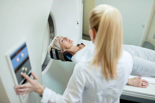
![Radiologist preparing patient for PET-CT scanner. View of radiologist or technician with senior male patient through an integrated PET-CT scanner.
[url=file_closeup.php?id=16785279][img]file_thumbview_approve.php?size=1&id=16785279[/img][/url] [url=file_closeup.php?id=17407688][img]file_thumbview_approve.php?size=1&id=17407688[/img][/url] [url=file_closeup.php?id=17132658][img]file_thumbview_approve.php?size=1&id=17132658[/img][/url]
[url=file_closeup.php?id=16785241][img]file_thumbview_approve.php?size=1&id=16785241[/img][/url] [url=file_closeup.php?id=16785267][img]file_thumbview_approve.php?size=1&id=16785267[/img][/url] [url=file_closeup.php?id=16802071][img]file_thumbview_approve.php?size=1&id=16802071[/img][/url]
[url=http://www.istockphoto.com/file_search.php?action=file&lightboxID=10736333]More PET-CT scanner and x-ray images[/url]
More medical imaging equipment:
[url=file_closeup.php?id=16727579][img]file_thumbview_approve.php?size=1&id=16727579[/img][/url] [url=file_closeup.php?id=16678574][img]file_thumbview_approve.php?size=1&id=16678574[/img][/url] [url=file_closeup.php?id=16727607][img]file_thumbview_approve.php?size=1&id=16727607[/img][/url]
[url=http://www.istockphoto.com/file_search.php?action=file&lightboxID=10654378]Mammography, MRI, CT scans[/url] pet scan stock pictures, royalty-free photos & images](https://media.istockphoto.com/id/155009721/photo/radiologist-preparing-patient-for-pet-ct-scanner.jpg?s=612x612&w=0&k=20&c=IJ2eWdTa7t0pi8qj4145YMSvClY2sUViX7BNy03P6GY=)
View of radiologist or technician with senior male patient through an integrated PET-CT scanner. [url=file_closeup.php?id=16785279][img]file_thumbview_approve.php?size=1&id=16785279[/img][/url] [url=file_closeup.php?id=17407688][img]file_thumbview_approve.php?size=1&id=17407688[/img][/url] [url=file_closeup.php?id=17132658][img]file_thumbview_approve.php?size=1&id=17132658[/img][/url] [url=file_closeup.php?id=16785241][img]file_thumbview_approve.php?size=1&id=16785241[/img][/url] [url=file_closeup.php?id=16785267][img]file_thumbview_approve.php?size=1&id=16785267[/img][/url] [url=file_closeup.php?id=16802071][img]file_thumbview_approve.php?size=1&id=16802071[/img][/url] [url=http://www.istockphoto.com/file_search.php?action=file&lightboxID=10736333]More PET-CT scanner and x-ray images[/url] More medical imaging equipment: [url=file_closeup.php?id=16727579][img]file_thumbview_approve.php?size=1&id=16727579[/img][/url] [url=file_closeup.php?id=16678574][img]file_thumbview_approve.php?size=1&id=16678574[/img][/url] [url=file_closeup.php?id=16727607][img]file_thumbview_approve.php?size=1&id=16727607[/img][/url] [url=http://www.istockphoto.com/file_search.php?action=file&lightboxID=10654378]Mammography, MRI, CT scans[/url]
Veterinary flat icons. Vector illustration include icon - stethoscope, grooming, , xray, ultrasound, vaccination, sterilization glyph silhouette pictogram for vet clinic. Black color.

Human brain with abstract visualization of neuron activity
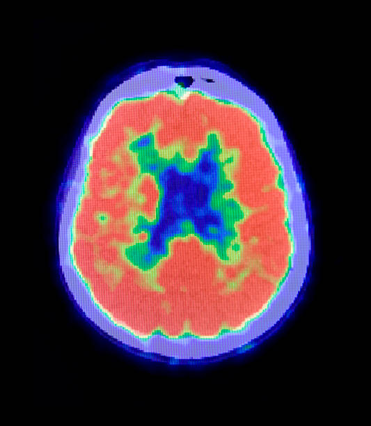
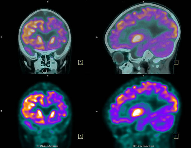
These are computer generated images, fusing PET (positron emission tomography) with CT (computed tomography) scan of the brain (in axial or horizontal cuts). It consists of injecting a radioactive analogue of glucose, FDG (fluorodeoxyglucose) into the bloodstream; the three-dimensional images of tracer concentration within the brain are then constructed by computer analysis. The more metabolically active areas will retain more FDG, and consequently retain more radiation (orange color). It is an important tool for detecting malignant tumor like metastasis mainly in other parts of the body. In the brain, it also has been used to detect areas of the brain that generate seizures.
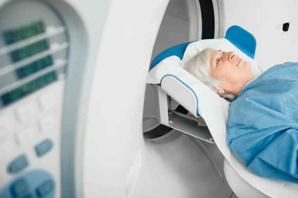
High angle view of mature woman and radiologist during MRI scan examination at clinic.
A black silhouette of a paw print, often used as a symbol for pets, wildlife, or animal-related themes.
Shot of a senior woman being comforted by a doctor before and MRI scanhttp://195.154.178.81/DATA/i_collage/pu/shoots/806398.jpg
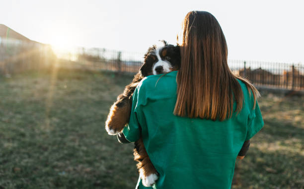
Young female veterinarian checking or examining purebred Bernese Mountain dog puppies.
![Woman Receiving a medical Scan for Breast Cancer Diagnosis Woman Receiving a medical Scan for Breast Cancer Diagnosis
[url=http://www.istockphoto.com/file_search.php?action=file&lightboxID=6833324] [img]http://www.kostich.com/cancer.jpg[/img][/url]
[url=http://www.istockphoto.com/file_search.php?action=file&lightboxID=4063973] [img]http://www.kostich.com/imaging.jpg[/img][/url] pet scan stock pictures, royalty-free photos & images](https://media.istockphoto.com/id/108268588/photo/woman-receiving-a-medical-scan-for-breast-cancer-diagnosis.jpg?s=612x612&w=0&k=20&c=y0gBwyhHdRLi4P6lGh2jO2l7WRKJBz5DIxe5h3cB_Uo=)
Woman Receiving a medical Scan for Breast Cancer Diagnosis [url=http://www.istockphoto.com/file_search.php?action=file&lightboxID=6833324] [img]http://www.kostich.com/cancer.jpg[/img][/url] [url=http://www.istockphoto.com/file_search.php?action=file&lightboxID=4063973] [img]http://www.kostich.com/imaging.jpg[/img][/url]
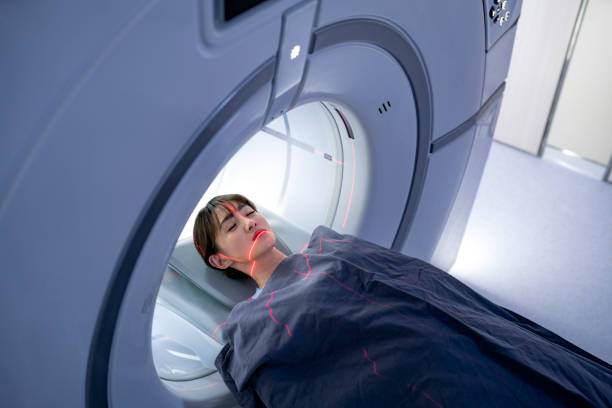
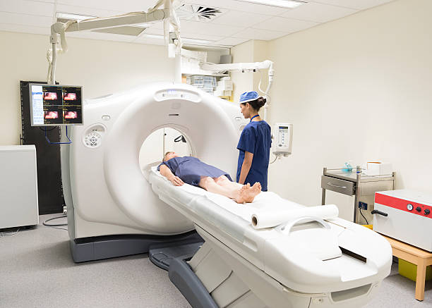
High angle view of patient lying for MRI scan. Sick young woman is going through medical procedure. She is with eyes closed.
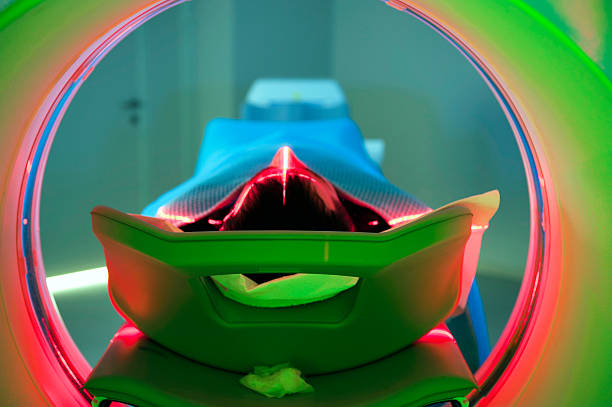
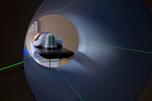
"Health series:Tomography Tunnel-PET/CT, Rear ViewTo see my other photos please click here:"
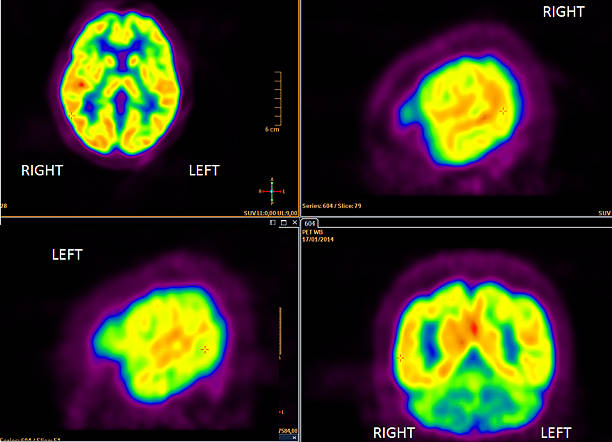
These are computer generated images called PET (positron emission tomography) of the brain. It consists of injecting a radioactive analogue of glucose, FDG (fluorodeoxyglucose) into the bloodstream; the three-dimensional images of tracer concentration within the brain are then constructed by computer computer analysis. The more metabolically active areas will retain more FDG, and consequently retain more radioation (orange color). It is an important tool for detecting malignant tumors such as metastasis mainly in other parts of the body. In the brain, it also has been used to detect areas of the brain that generate seizures, they are less metabolically active.
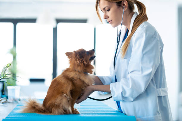
Shot of beautiful young veterinarian woman using stethoscope to listening to the heartbeat of cute lovely pomeranian dog at veterinary clinic.

Blank Cat Dog Id Tag Name Pendant Necklace Collar Mockup, 3d render illustration.
Set is designed with suitable visuals for all medical and healthcare

A brown rabbit sits in the grass in a clearing on a summer day. The rabbit is sitting in a basket. The girl's hands caress the rabbit.
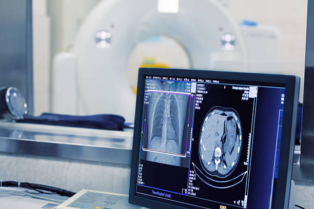
Medical CT or MRI or PET Scan Standing in the Modern Hospital Laboratory room in hospital, Medical Equipment and Health Care
Woman in having CAT scan with radiologist by machine. PET scan equipment. Medical scan of patient. Radiography.

Radiologist getting a patient ready for an MRI at the hospital - medical exam concepts
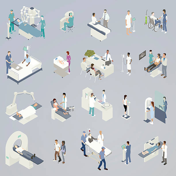
20 spot illustrations of medical procedures and related icons, presented in isometric view and in a flat, consistent color palette. Includes: robot-assisted surgery, medical consultations, checking blood pressure, attending to a newborn, sonogram, non-invasive radiation treatment, dental visit, blood or pharmaceutical lab analysis, weight scale during checkup, mammogram, MRI/CT scan/Pet scan, physical therapy, an injured man with neck brace in gurney with paramedics, and a woman receiving an x-ray.

Patient getting a CAT scan at the hospital - healthcare and medicine concepts

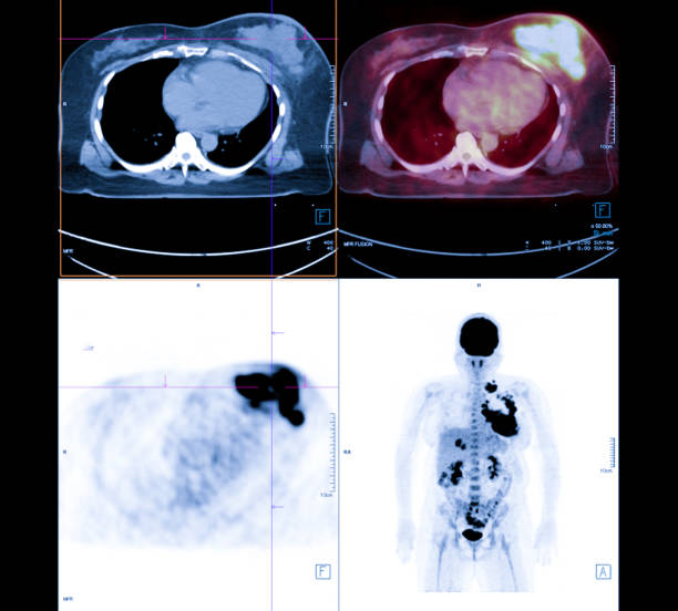
PET scan fusion MRI prostate gland fusion imaging in high-grade prostate cancer.

"Man Receiving Scan,Tomography Tunnel-PET/CTTo see my other photos please click here:"
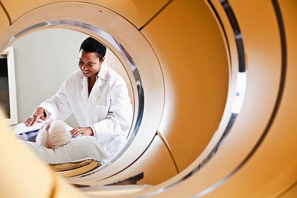
View of radiologist or technician with senior male patient through an integrated PET-CT scanner.

Veterinarians doctors in blue uniforms conduct a routine examination of a dog on a table in a modern office of a veterinary clinic. Treatment and vaccination of pets.
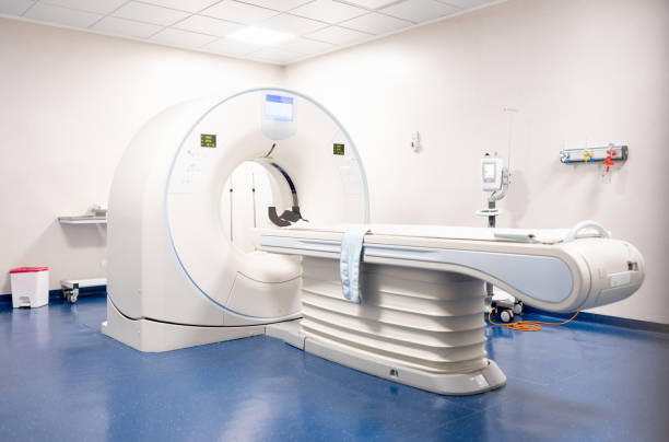
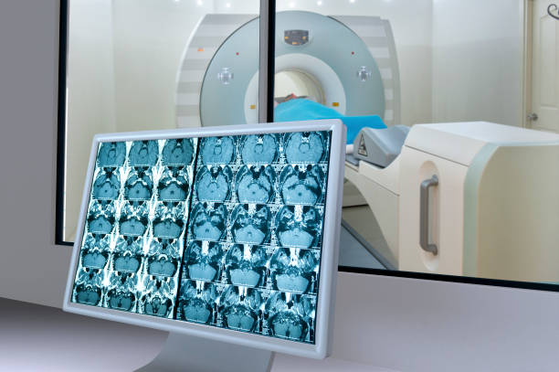
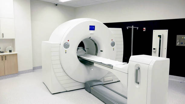
MRI scanner in an empty hospital room - healthcare and medicine concepts

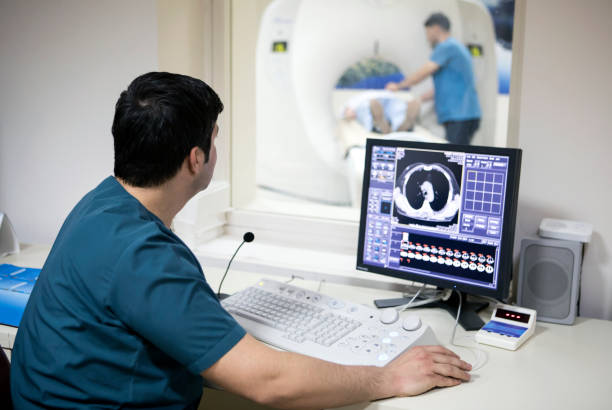
Radiologist in the control room performing a medical scan - healthcare and medicine concepts
Recycling code 1 (PET - Polyethylene terephthalate) outline icons set. Empty clear plastic bottles on white background.

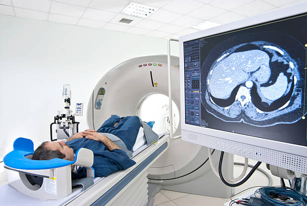
Patient lying down getting a CAT scan at the hospital - healthcare and medicine concepts
http://cginspiration.com//Istock/V2/WhiteCharacters.jpg
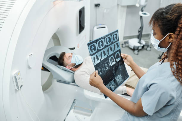
Young African radiologist in uniform and protective mask looking at x-ray image of patient lying on long couchette of medical equipment

These are computer generated images called PET (positron emission tomography) of the brain. It consists of injecting a radioactive analogue of glucose, FDG (fluorodeoxyglucose) into the venous circulation; the three-dimensional images of tracer concentration within the brain are then constructed by computer analysis. The more metabolically active areas will retain more FDG, and consequently retain more radiation (orange color). It is an important tool for detecting malignant tumor like metastasis mainly in other parts of the body. In the brain, it also has been used to detect areas of the brain that generate seizures.
A set of veterinarian icons with editable strokes or outlines using the EPS file. The icons include a dog with a cast on its broken leg, dogs paw in a vets hand, pet health care, veterinarian with hand on puppy dog, pet immunization, cat getting ears checked at vet, German Shepard dog, veterinarian giving a dog a medical examination in office, pet nail clipping, vet holding up an x-ray of a pets broken bone, vet using a stethoscope to examine a dog in his care, hand shielding a puppy dog, dog with a heart in the background, vet giving owner his dog after veterinarian check-up, veterinarian with stethoscope standing next to her assistant, vet medical checklist, dog with identification chip, Labrador retriever, dog getting eye exam by veterinarian, pet medication, pet surgery, veterinarian clinic, dog getting its ear cleaned and a couple holding their pet dog after vet visit.